The Process Of Movement Between The Brain And Body

Julia C. Basso, PhD
Did you ever wonder how we move?
The ability to move is an interconnected process between the body and brain. As I have discussed before, the motivation to move is regulated by the reward circuitry of our brain. But what actually happens when the brain gets the signal, “ok, let’s get moving”? There are several levels to this complicated process.
The Several Levels
First, information from the brain, a cortical region known as the primary motor cortex, needs to travel to the muscles. It does this by traveling through a pathway known as the pyramidal system. The pyramidal system consists of neurons traveling from the primary motor cortex to an area of the brain stem known as the medulla. At this point, the neurons cross over, from right to left and right to left. This is why your right side of the brain controls the left side of the body and vice versa. From here, the neurons travel down the spinal cord and out to the muscles.
At the point where the neuron ends and the muscle begins, there is a space that is known as the neuromuscular junction. When the action potential reaches the end of the neuron, which is carrying information from the brain to the muscle, a neurotransmitter called acetylcholine is released. This causes an electrical signal in the muscle, which triggers the muscle to contract. The muscular contraction is an actual physical process of filaments within the muscle, known as actin and myosin, coming together.
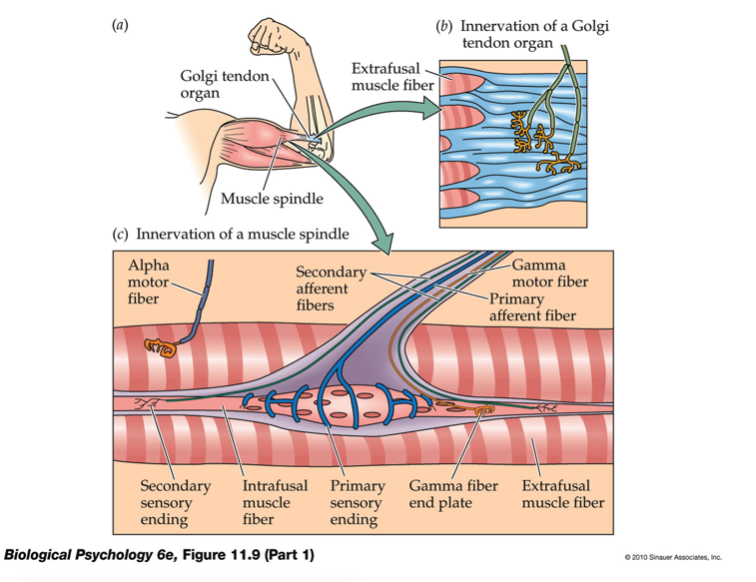
To continue moving, of course, the brain needs to be informed of what just happened. Within the muscle, two types of receptors exist – golgi tendon organs and muscle spindles. The golgi tendon organs sense force or tension in a muscle. Neurons are attached to these receptors, which then send information back to the brain that the muscle is contracted. Muscle spindles, on the other hand, sense the length of the muscle. Neurons connected to these receptors signal to the brain that the muscle is stretched.
Muscle Fibers
 Two types of muscle fibers exist, which help us to perform all of the activities we do throughout the day. One type is the namesake of this website – fast twitch muscles; the other is slow twitch muscles.
Fast twitch muscles contract rapidly but fatigue easily. They are useful for when we want to perform powerful, rapid activities like lifting a heavy weight or sprinting. Slow twitch muscles contract much more slowly but are resistant to fatigue. These muscles help with postural balance as well as endurance exercises like jogging or running.
Positive Effects On The Brain
As a neuroscientist who studies the effects of exercise on the brain, I know that exercise has profound effects on brain plasticity – or the ability of the brain to change in positive ways. What I didn’t know, though, was whether or not exercise affected the motor system down to the level of the neuromuscular junction.
It turns out that the answer is overwhelmingly, yes. For instance, exercise significantly increases the size of the motor cortex. Endurance training also increases the branching of motor neurons, causing there to be more connections between the neurons and the muscle (Deschenes et al., 2016). In addition, long-term exercise causes an increase in the amount of acetylcholine as well as the number of acetylcholine receptors, making the neuromuscular junction more effective.
Takeaway
The motor system is a complicated network of neurons, starting from the brain and ending at the muscle. Interestingly but not totally surprisingly, the motor system benefits from exercise at all of its many levels. Now that you know how the body moves, go get moving!
Related Article: Get Motivated To Exercise: Rats Do It, You Can Too
References:
Deschenes, M. R., Kressin, K. A., Garratt, R. N., Leathrum, C. M., & Shaffrey, E. C. (2016). Effects of exercise training on neuromuscular junction morphology and pre-to post-synaptic coupling in young and aged rats. Neuroscience, 316, 167-177.
You Might Like:


